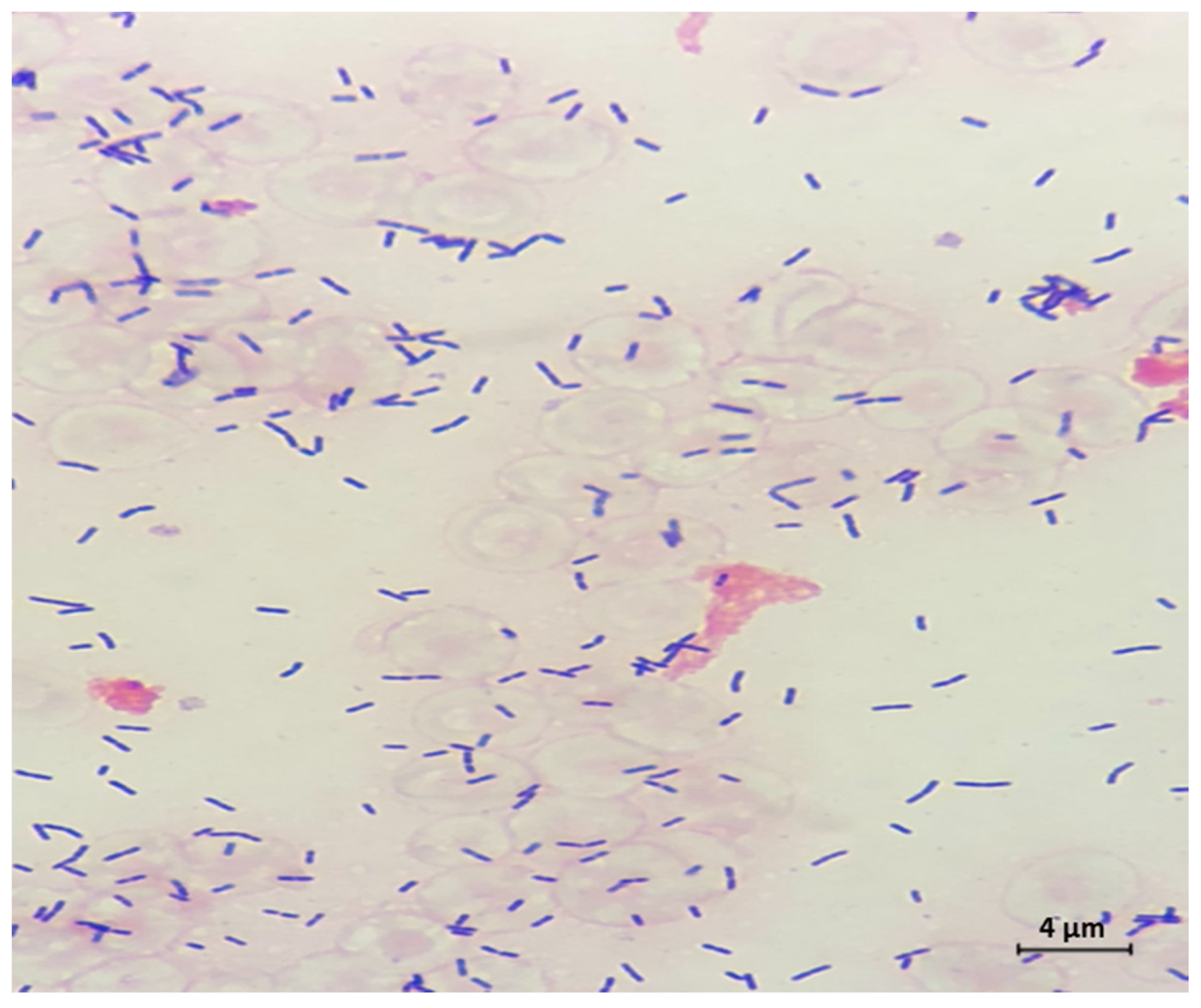
Listeria Monocytogenes Gram Stain Morphology
Possess a stable morphology. Cocci Clusters Staph Species Coagulase Positive.

Bacteria 101 Cell Walls Gram Staining Common Pathogens Tusom Pharmwiki
The gram stain was devised by histologist Christian Gram as a method of.
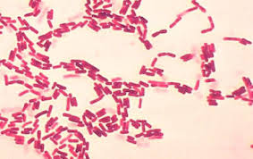
Listeria monocytogenes gram stain morphology. Morphology cocci bacilli coccobacilli spiral or presence of branching filaments Gram-staining properties Grampositive Gramnegative and atypical metabolic activity aerobic anaerobic microaerophile or facultative or virulence factors eg presence of capsule pili proteins formation. Several different serogroups of Shigella are described. Listeria monocytogenes our previous study 14 Pseudomonas fluorescens 15 Paeruginosa 16 Salmonella senftenberg 17 and Sputrefaciens 18 etc.
It was discovered by a German physician Robert Koch in 1876 and became the first bacterium to be. Gram Stain Mechanism. Bacillus anthracis is a Gram-positive and rod-shaped bacterium that causes anthrax a deadly disease to livestock and occasionally to humansIt is the only permanent pathogen within the genus BacillusIts infection is a type of zoonosis as it is transmitted from animals to humans.
You see what looks like cocci and short bacilli. ε-Polylysine ε-PL is a safe food additive that is used in the food industry globally. Salmonella enterica serovar Typhimurium phagosomes also are diverted from the normal endocytic pathway of phagosome-lysosoma fusion 42 233 and this bacterium requires acidification of the phagosome to survive in macrophages 234.
The results showed that minimum inhibitory concentrations MICs ranged between 0. However the Gram stain result is critical to diagnosis and how to interpret a Gram stain result should still be covered in a nursing prerequisite microbiology course albeit with less emphasis on how to. Gram-positive bacteria retain the color of the primary stain crystal violet in the Gram staining procedure and appear as purpleviolet under a light microscope.
Listeria monocytogenes is a human pathogen. Gram-positive bacteria stain purple while the Gram-negative bacteria stain Pink after losing the purple color during the alcohol was thus taking up the safranin. However the application of Linalool or essential oils is chal-.
Feb 16 2021 Rugose colony morphology is usually related to high levels of c-di-GMP. It is generally recognized as safe GRAS as a food additive. Gram-_____ cells stain purple whereas gram-_____ cells stain pink or red when using the Gram stain technique.
Academiaedu is a platform for academics to share research papers. Gram staining has been especially used because of its ability to differentiate bacteria base on their cell wall content a major characteristic that classifies bacteria into two types namely Gram-positive and Gram-negative bacteria. As the vast majority of nurses 983 do not perform and read Gram stains it is unsurprising that the nurses assigned the lowest relevance to Gram stain procedures and microscope use.
Flexneri belongs to group B. What is the most likely morphology of these microorganisms. For example in this case it is not clear how to determine the colony size.
Gram positive cocci in pairs and chains may suggest Streptococcus species or Enterococcus species Branching Gram positive rods modified acid fast stain positive may suggest Nocardia or Streptomyces species. Tumbling motility with Listeria monocytogenes. This kind of growth can be difficult to describe using all of the bacterial colony morphology characteristics.
Gram Stain 146 Staining Properties of Gram-Positive and Gram-Negative Bacteria 147 Types of Media 148 Routine Media for Aerobes and Facultative Anaerobes 149 Selective Media for Isolation of Neisseria gonorrhoeae and Neisseria meningitidis 152 Special Bacteriologic Media 153 Aerotolerance Test 155 xiii Organisms Requiring Incubation in. Gram Positive Cell Wall. Last updated on May 26th 2021.
This study evaluated the antimicrobial and antibiofilm activity of antibacterial peptides ε-PL against food poisoning pathogens detected in chicken Salmonella Enteritidis Listeria monocytogenes and Escherichia coli. Shigella flexneri is a species of Gram-negative bacteria in the genus Shigella that can cause diarrhea in humans. Listeria and Shigella physically escape the phagosome and replicate in the cytoplasm and Legionella inhibits phagosome-lysosome fusion.
Human pathogenic bacteria can be classified according to their characteristics. Flexneri infections can usually be treated with antibiotics although some strains have become resistantLess severe cases are not usually treated because they become more resistant in the. Gram positive cocci in clusters may suggest Staphyloccocus species.
These bacteria have a cell wall containing a thick layer of peptidoglycanOn the basis of cell morphology Gram-positive bacteria are divided mainly into two groups Gram-positive cocci and. Gram-positive bacteria have a thick mesh-like cell wall which is made up of peptidoglycan 50-90 of cell wall which stains purple.

Colony Morphology And Gram Staining Property Of The Isolated Lab From Download Scientific Diagram
Gram Staining Rules

Pin On Microscopio

Mixed Culture Art Print Watercolor Science Medical Science Art Biology Science Gift Microbiology Biology Art Laboratory Prints Biology Art Microbiology Science Gifts

Pin On Escherichia Coli Gram Stain

Pin On Bacteriology

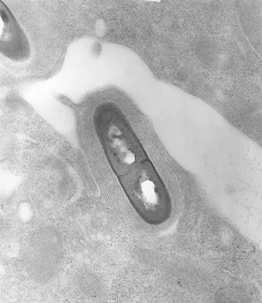
Gram Stain Of L Monocytogenes Mackaness A And L Monocytogenes Riii Download Scientific Diagram

Gram Staining Of Selected Bacteria A L1 Strain And B L2 Strain Download Scientific Diagram
Genus Listeria Definition Class Characteristics And Gram Stain

Microsporum Gypseum Medical Laboratory Science Microbiology Medical Laboratory

Pdf Studies On The Isolation Of Listeria Monocytogenes From Food Water And Animal Droppings Environmental Health Perspective

Pseudomonas Putida Kids Rugs Taxonomy Microbiology

Gram Staining Of C Ramosum Demonstrating Thin Long Gram Negative And Download Scientific Diagram

Gram Staining Morphological Observation Of Salmonella Typhimurium And Download Scientific Diagram

Colony Morphology And Gram Staining Property Of The Isolated Lab From Download Scientific Diagram
Ijerph Free Full Text Double Negative T Cell Reaction In A Case Of Listeria Meningitis Html

Gram Stain Qc Bacteria Bacteria Art Science Science Art Etsy Science Art Science Gifts Biology Art

Gram Staining Morphological Observation Of Salmonella Typhimurium And Download Scientific Diagram

Pin Su Scienza Arte